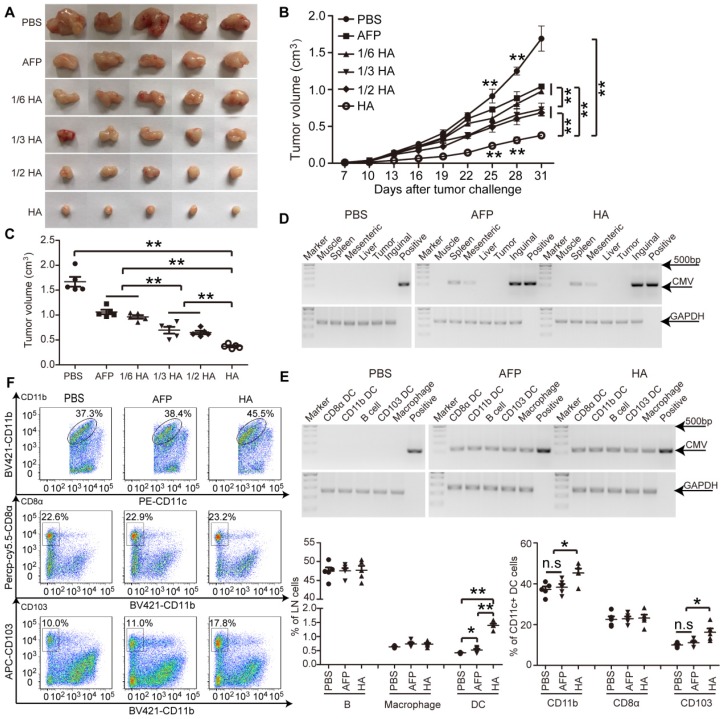
Figure 2.
Evaluation of efficacy and homing capacity of lenti-HA in ectopic HCC mice. Different amounts of lentivirus were subcutaneously administered into day-7 ectopic HCC mice for 3 weeks at weekly interval. (A) Representative tumor images for treated mice. (B-C) Analysis of tumor volume from ectopic HCC mice treated with different doses of lenti-HA or lenti-AFP at different time-points (n=5, **P<0.01). Tumors were harvested at 31 days after tumor challenge in (C). Semi-quantitative PCR results for showing the tissue distribution (D) or cellular localization (E) of lentivirus in ectopic HCC mice 3 days after last injection. The plasmid was used as a positive control. (F) Flow cytometric analysis of immune cells in lymph nodes (n=5, *P<0.05; **P<0.01). N.s refers to not significant. Two-tailed t test was used for statistical analysis and all experiments were repeated twice (two repeated experiments yielded similar results and thus one representative result was shown).

