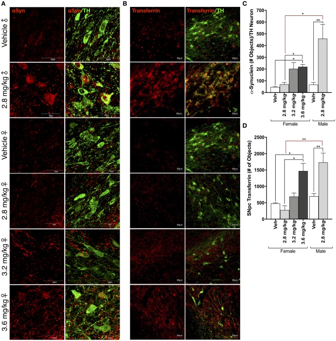
Figure 4.
Sex differences in protein accumulation following rotenone treatment. A, Representative confocal microscopy (×100) of α-synuclein (αSyn, red) within tyrosine hydroxylase (TH)-positive neurons (green) within the substantia nigra (SN) in male and female rats following vehicle or rotenone treatment. B, Representative images (×20 montage) of transferrin (Tf; red) accumulation within tyrosine hydroxylase TH-positive neurons (green) within the SN in male and female rats following vehicle or rotenone treatment. C, Quantification of α-synuclein within TH-immunoreactive cells within the SN (F(1, 15) = 15.12, p = .0015; two-way analysis of variance [ANOVA]). D, Quantification of transferrin within TH-immunoreactive cells within the SN (F(1, 14) = 24.45, p = .0002; two-way ANOVA). (For interpretation of the references to colour in this figure legend, the reader is referred to the web version of this article.)

