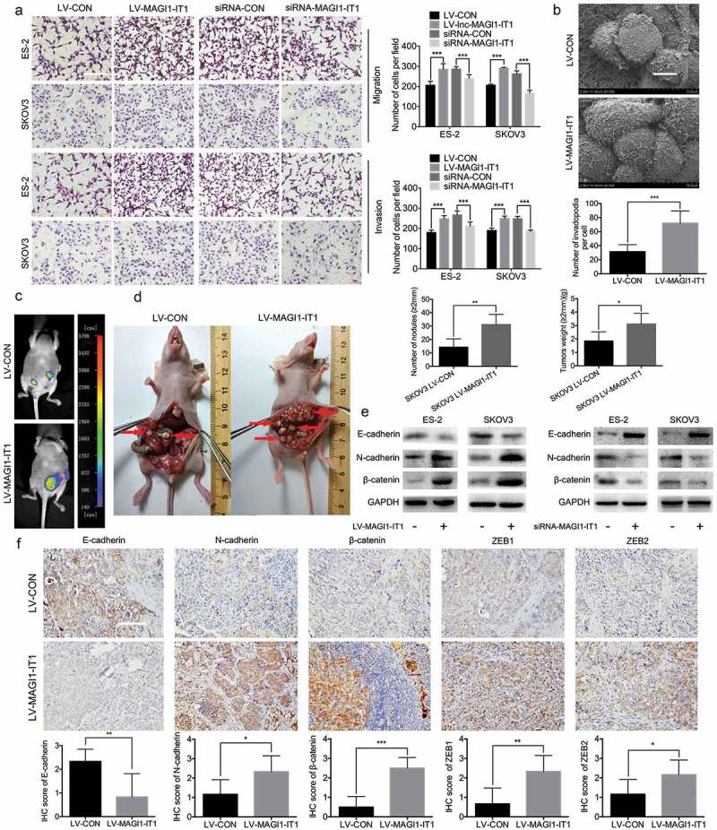
Figure 3.

MAGI1-IT1 markedly enhanced EOC cell invasion and metastasis in vitro and in vivo.
A. Transwell assays of dysregulation of MAGI1-IT1 in EOC cells in vitro. Scale bar, 100 μm. B. SEM and statistical numbers of invadopodia in MAGI1-IT1-overexpressing SKOV3 cells. Scale bar, 50 μm. C. The growth of MAGI1-IT1-overexpressing SKOV3 orthotopic ovarian xenografts was detected with a NightOWL LB 983 In Vivo Imaging System. D. After sacrifice, the ovarian tumors in nude mice were removed and are shown by red arrows in the images. The average number of peritoneal tumor nodules and average weight of tumors from each group were quantified. E. Western blot analysis of EMT markers in dysregulation MAGI1-IT1 EOC cells. F. Representative IHC staining and average scores for EMT markers in orthotopic ovarian xenografts. Scale bar, 50 μm. All data were analyzed using Student’s t-test and are expressed as the mean ± SD, and statistically significant differences are presented as follows: *P < 0.05, **P < 0.01 and ***P < 0.001.
