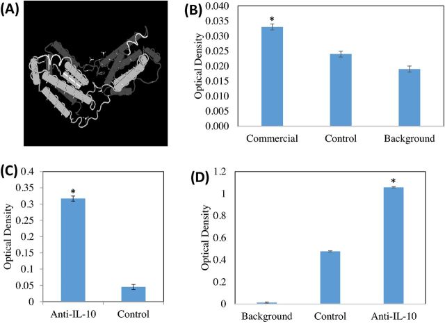
Figure 1.
Anti-IL-10 antibody binds to commercial recombinant IL-10 as well as IL-10 in the lumen of the small intestine. (A) A three-dimensional structure of human IL-10 (2H24_A, pubmed.gov) was used in combination with sequence alignment to determine the placement of the peptide used to bind to chicken IL-10. The region shown in white represented the 8 amino acid peptide used in this study. (B) Commercial rabbit anti-IL-10 antibody was shown to bind commercial recombinant chicken IL-10. This study was conducted to determine the usefulness of commercial rabbit anti-IL-10 as a chicken IL-10 capture antibody. In this experiment, commercial recombinant chicken IL-10 was bound to the plate, and the ability of commercial rabbit anti-IL-10 (commercial), rabbit isotype control (control), or the absence of any primary antibody (background) was compared (secondary antibodies used were goat anti-rabbit IgG-HRP conjugates). *Denotes different binding of commercial recombinant chicken IL-10 by the rabbit anti-IL-10 antibody and the isotype control rabbit antibody and the primary antibody blank (P < 0.05). (C) Commercial recombinant chicken IL-10 was coated on an ELISA plate and control or affinity-purified peptide specific antibody to IL-10 was used to demonstrate the specificity of the antibody in binding recombinant chicken IL-10. We demonstrate that the affinity-purified peptide antibody binds the recombinant IL-10 significantly over control antibody (P < 0.05). (D) IL-10 from intestinal contents of the duodenum of chickens was demonstrated using a capture ELISA (P < 0.05). In this ELISA, commercial rabbit anti-IL-10 was coated on the plate. Duodenal fluids from Eimeria spp.-challenged chicks were incubated on the bound rabbit anti-IL-10. Next incubation was either an affinity-purified chicken anti-IL-10 antibody (anti-IL-10) or control antibody (Control) or the secondary antibody alone (Background). Using an ELISA, IL-10 in the duodenal luminal fluid was qualitatively demonstrated in the duodenal luminal fluid of Eimeria-challenged chicks.

