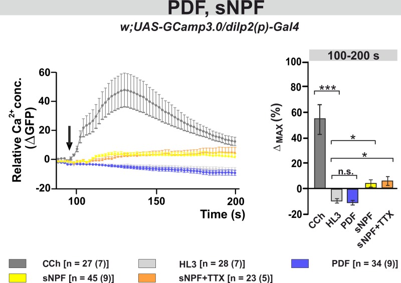
Fig 8. The neuropeptide sNPF induces a small increase in the intracellular Ca2+ level, while PDF has no effect.
Left panel: Average changes in GFP fluorescence of IPCs reflecting intracellular changes in Ca2+ levels. Substances were bath-applied to freshly dissected fly brains at ~100 s (indicated by a black arrow). The cholinergic agonist carbamylcholine (1 mM CCh) was used as positive control, which induced a robust increase in Ca2+, indicating that the general procedure was working. As a negative control, hemolymph-like saline (HL3) was applied. Application of 10–5 M synthetic PDF peptide did not alter Ca2+ levels, while 10–5 M sNPF induced a small but significant increase in Ca2+ levels. This calcium response seems to be due to direct activation of the IPCs, since it was not blocked in the presence of the sodium channel blocker TTX (2μM). Right panel: Maximum Ca2+ changes (%) for each individual neuron were calculated and averaged for each pharmacological treatment from 100 s until 200 s. Statistical comparison revealed significant increases in Ca2+ levels compared to the negative control (HL3) for CCh, sNPF and sNPF+TTX, while PDF had no effect. Data are presented as mean ± SEM. The color code of the different treatments and the number of neurons [n] and dissected brains (n) considered in this analysis are shown below the panels. Kruskal-Wallis test followed by Bonferroni-corrected Wilcoxon pairwise-comparisons. ***p<0.001, *p<0.05, n.s. not significant.

