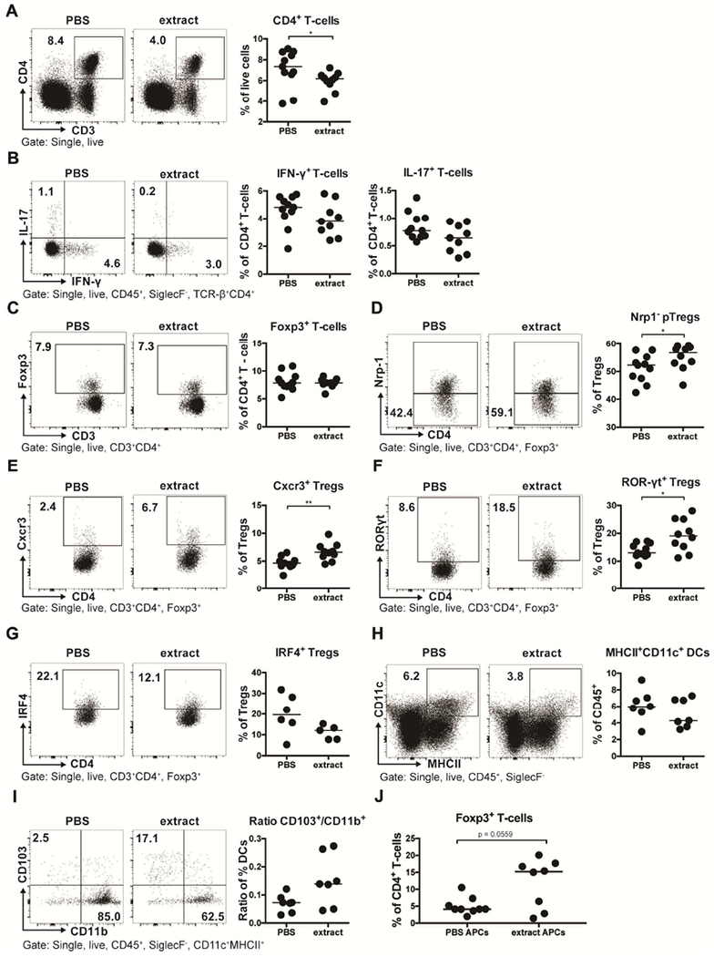
Figure 3. Perinatal trans-maternal exposure to H. pylori extract skews lung T-cell responses towards specific regulatory T-cell subsets.

(A-I) Mice were trans-maternally pre- and postnatally exposed to H. pylori extract or PBS during pregnancy and lactation. At six weeks of age, lung leukocytes were analyzed by multi-color flow cytometry. (A) CD4+ T-cell frequencies among all leukocytes; representative FACS plots are shown on the left. (B) Th1 and Th17 frequencies among all CD4+ T cells, of the mice shown in A. (C) Foxp3+ Treg frequencies of the same mice. (D) Peripherally induced Treg (pTreg; Nrp-1−) frequencies among all Foxp3+ Tregs. (E-G) Frequencies of Cxcr3+, RORγT+ and IRF4+ Tregs among all CD4+FoxP3+ Tregs. (H) MHCII+CD11c+ dendritic cell (DC) frequencies among all pulmonary leukocytes. (I) Ratios of CD103+CD11b− over CD103−CD11b+ DCs, of all mice shown in H; representative FACS plots are shown on the left. (J) Frequencies of Foxp3+ T-cells among all CD4+ OT-II T-cells, after three days of co-culture with FACS-sorted splenic MHCII+CD11c+ DCs. Data are pooled from two studies (A-F) or show the results of a representative experiment of at least two (G-J). Horizontal lines indicate medians; an unpaired Mann-Whitney U test was used throughout. * p<0.05, ** p<0.01.
