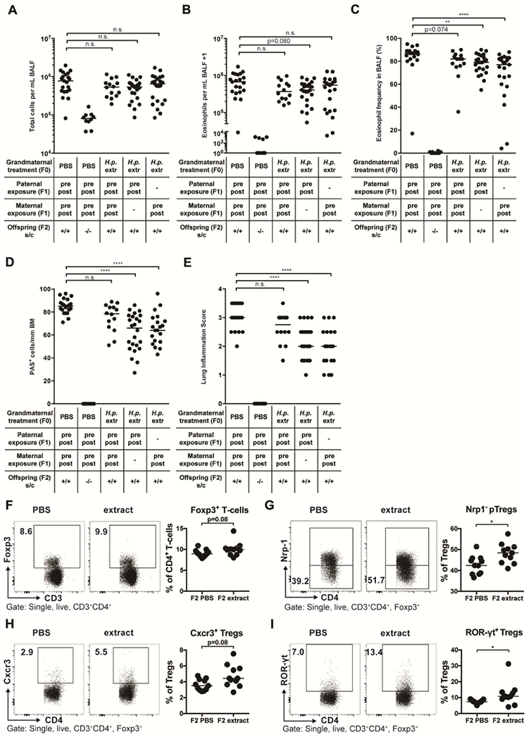
Figure 7. H. pylori extract induces inter-generational protection against allergic airway inflammation and skews lung T-cell responses of the F2 generation.

(A-I) F0 dams were subjected to twice-weekly oral gavage with H. pylori extract (H.p. extr) or PBS during pregnancy and lactation. Perinatally exposed F1 animals obtained in this manner were bred with each other or with naïve mates. At six weeks of age, F2 progeny were subjected to flow-cytometric analysis of the pulmonary T-cell compartment (F-I) or sensitized and challenged intranasally with house dust mite (HDM) allergen (A-E). (A) Total leukocytes in 1 ml of BAL fluid (BALF). (B) Total eosinophils in 1 ml of BALF. (C) Eosinophil frequencies in BALF. (D,E) Pulmonary inflammation and goblet cell metaplasia. (F) Foxp3+ Treg frequencies among all T cells. (G) Peripherally induced Treg (pTreg; Nrp-1−) frequencies among all Foxp3+ Tregs. (H,I) Frequencies of Cxcr3+ and RORγt+ Tregs among all CD4+FoxP3+ Tregs. In A-I each symbol represents one mouse. Results were pooled from three (A-E) or two (F-I) independent experiments. In A-E, ANOVA with Dunn’s multiple comparison correction was used for calculation of p-values. In F-I, an unpaired Mann-Whitney U test was used for calculation of p-values. * p<0.05, ** p<0.01, **** p<0.0001.
