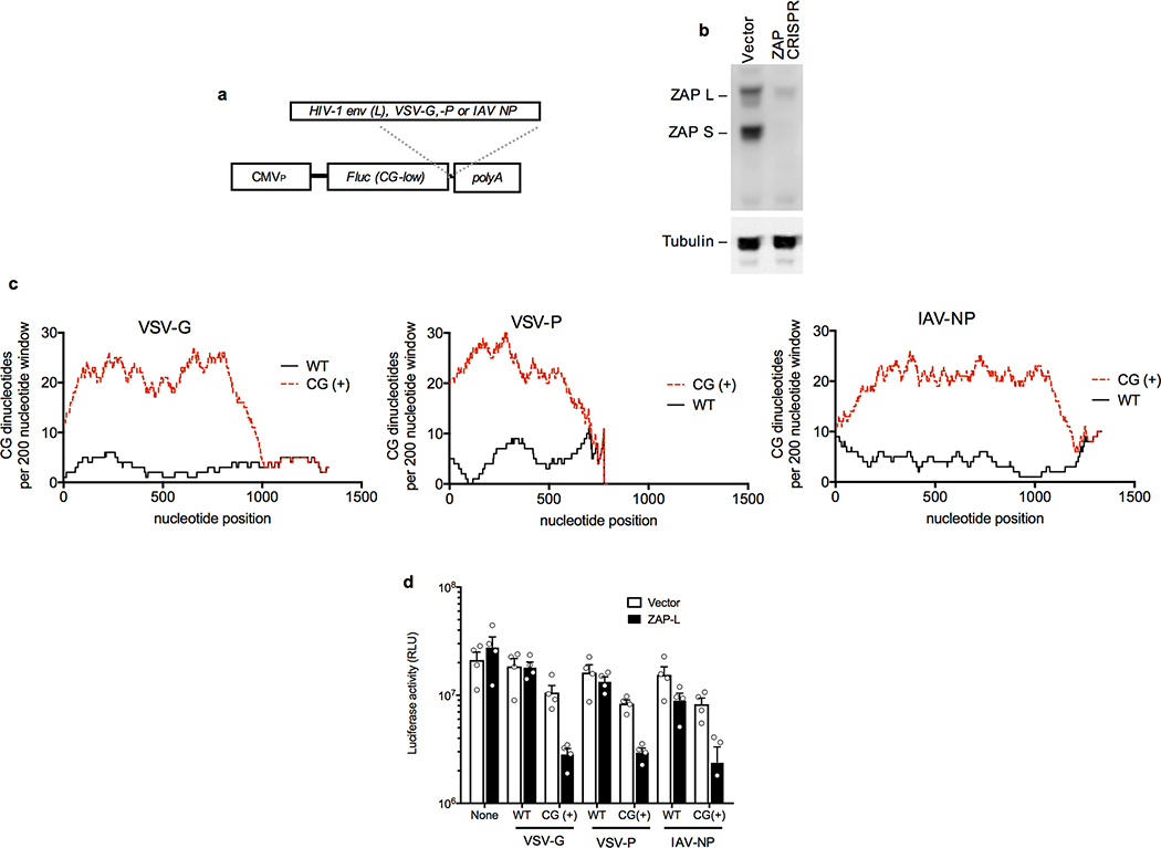
Extended data Figure 6. CG dinucleotides in 3′UTRs confer sensitivity to inhibition by ZAP.
a, Schematic representation of a reporter construct encoding a CG-dinucleotide depleted fluc cDNA into which were inserted the indicated sequences as 3′UTRs. b, Western blot analysis of ZAP expression following CRISPR mutation of ZAP exon 1 in HeLa cells. Representative of 3 experiments. c, Number of CG dinucleotides present in a 200-nucleotide sliding window in the indicated viral cDNA sequences that were left unmanipulated (WT), or recoded with synonymous mutations to contain the maximum number of CG dinucleotides (CG+). d, Luciferase expression following transfection of 293T ZAP−/− cells with CG-dinucleotide depleted fluc reporter plasmids incorporating the indicated VSV or influenza A virus (IAV) RNA sequences as 3′UTRs, in the presence or absence of a cotransfected ZAP-L expression plasmid (mean ± sem n=4 independent experiments).

