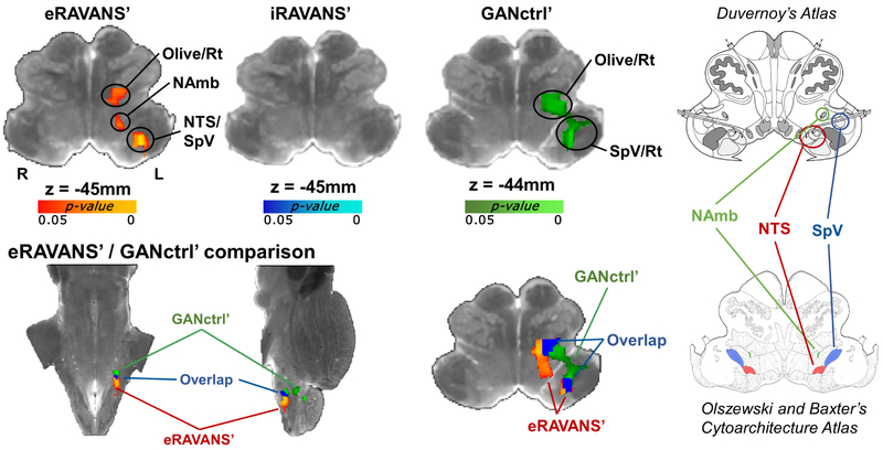
Figure 3 –
Top row: group maps showing masked ipsilateral medullary responses to exhalatory taVNS (eRAVANS’), inhalatory taVNS (iRAVANS’) and greater auricular nerve control stimulation over the earlobe (GANctrl’), overlaid on a high-resolution (0.2 mm) ex vivo brainstem. The respiration phase-matched sham (eSham, iSham) was subtracted from each active stimulation condition in order to normalize active stimulation response and control for respiratory modulation of the fMRI signal. Bottom row: eRAVANS’ (red-yellow) and GANctrl’ (green) group maps, as well as their overlap (blue), are shown on the same underlay. The corresponding brainstem slices from the Duvernoy’s (top right) and the Olszewski and Baxter’s (bottom right) atlases aid the localization of functional responses. The eRAVANS’ cluster is consistent with purported NAmb, NTS and part of SpV, whereas the GANctrl’ response mainly involves a cluster more consistent with SpV.

