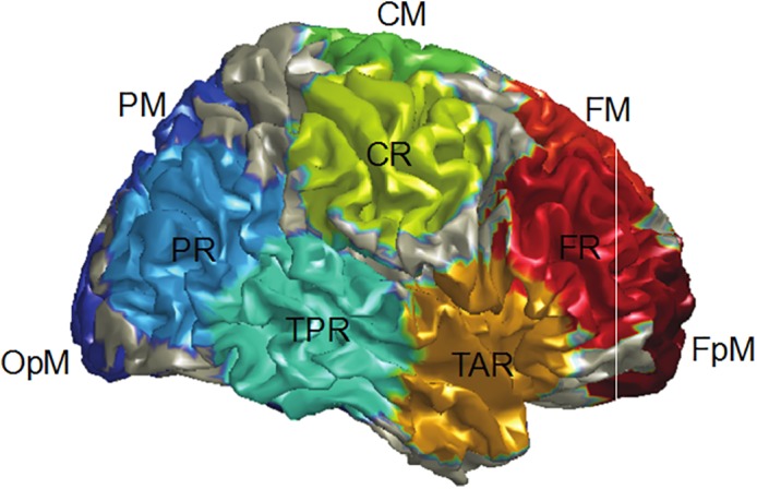
Figure 1.

Regional source locations. Resting‐state alpha activity was measured for all 15 sources, with the PAF for each participant determined via identifying from the 15 alpha power spectra the frequency at the source with the most alpha activity within a 7–13 Hz range (typically the midline parietal or occipital source). The figure shows the locations for FpM fronto‐polar midline, FM frontal midline, FR frontal right, TAR temporal anterior right, CM central midline, CR central right, TPR temporal posterior right, PM parietal midline, PR parietal right, and OPM occipito‐polar midline. The left hemisphere contained analogous regional sources, though with region source labels ending with “L” instead of “R” [Color figure can be viewed at http://wileyonlinelibrary.com]
