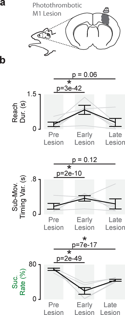
Figure 7. Persistent disruption of skilled fine movements after M1 lesion.
a. Illustration of photothrombotic M1 lesion. b. Differences in reach duration, sub-movement timing variability, and success rate between trials before M1 lesion (Pre Lesion), trials during the first reaching session post- lesion (Early Lesion), and trials once a performance plateau had been reached (Late Lesion; n = 5 animals). Grey lines represent mean values from individual animals and black lines represent mean and SEM across animals. P values from mixed-effects models.

