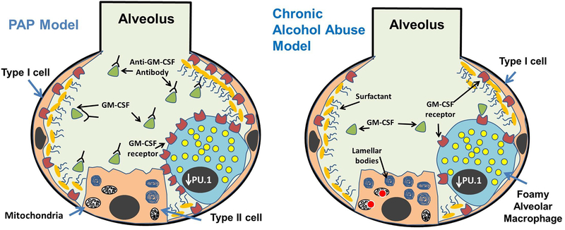
Figure 1:
Comparison of alveolar pathologies between pulmonary alveolar proteinosis (PAP) and chronically ethanol exposed lung. A hallmark of both conditions is the lipid-laden morphology of macrophages caused by the accumulation of lipids (yellow circles) in the cytosol, and the development of immunoregulatory dysfunction. Acquired forms of PAP develop because of autoantibodies preventing GM-CSF (green triangles) from binding to receptors in the plasma membranes of alveolar macrophages and type II alveolar epithelial cells. By contrast, levels of GM-CSF receptors (red color shapes) are reduced in the alveoli in response to chronic alcohol exposure. Lipid synthesis is also increased in alveolar epithelial type II cells in response to alcohol. In both the PAP and chronic alcohol exposed lung, decreased GM-CSF signaling leads to reduced PU.1 levels in the nucleus (charcoal gray oval) of AMs. Increased ROS levels have been observed in the mitochondria of ethanol metabolizing cells, which may also contribute to alterations in lipid homeostasis in the lung after chronic ethanol consumption.

