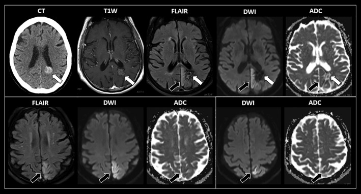
Figure 1.

Sequential axial imaging studies of this patient with recurrent abdominal motor seizures. Images inside a white square correspond to the same axial cut. An acute subcortical hemorrhage is evidenced in the left mesial parietal region (white arrows). There is adjacent cortical cytotoxic edema extending cranially through the mesial aspect of the left parietal lobe to reach the postcentral and central sulci near the vertex. This region corresponds to the somatotopic localization of the sensorimotor areas corresponding to the trunk (black arrows). MRI sequences: T1W, T1‐weighted MR with intravenous contrast; FLAIR, fluid‐attenuated inversion recovery; DWI, diffusion‐weighted imaging; ADC, apparent diffusion coefficient.
