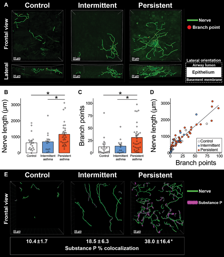
Fig. 1. Airway sensory innervation and substance P expression are increased in moderate persistent asthma.
(A) Representative 3D nerve models generated from bronchoscopic human airway biopsies immunolabeled with antibody to the pan-neuronal protein PGP9.5. (B and C) Bar graphs showing nerve length (B) and branch points (C) in samples derived from healthy subjects (control) and patients with mild intermittent asthma (intermittent) and moderate persistent asthma (persistent). (D) Correlation between nerve length and branch points in control and asthmatic patients (r2 = 0.87, P < 0.0001). (E) Representative 3D nerve models generated from bronchoscopic human airway biopsies obtained from healthy subjects (control) and patients with mild intermittent asthma (intermittent) and moderate persistent asthma (persistent), immunolabeled with antibody to neuronal substance P, and the pan-neuronal protein PGP9.5. Movies of nerve modeling are available in the online supplement (movies S1 to S3). Data are presented as means ± SEM. Asterisk (*) indicates P < 0.05 compared to all other groups. Statistical significance was determined using one-way analysis of variance (ANOVA) with a Bonferroni post hoc test (B, C, and E) and linear regression (D). In total, 63 subjects were included in the final analysis. Three biopsy specimens were analyzed per subject with 10 randomized z-stack images obtained per specimen.

