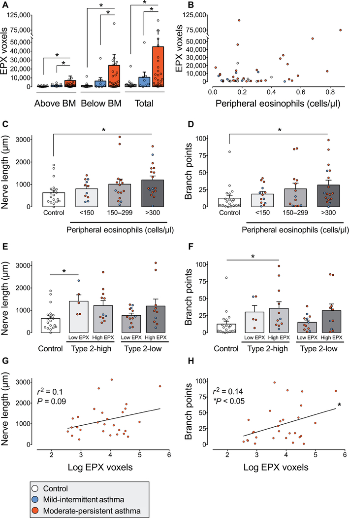
Fig. 2. Airway and peripheral blood eosinophils are associated with increased airway innervation in asthma.
(A) Eosinophil peroxidase (EPX) in human airway biopsies from healthy subjects (control, white bars) and from patients with mild intermittent (blue bars) and moderate persistent asthma (red bars). EPX was quantified above and below the epithelial basement membrane (BM). Data points represent the average of three biopsies per subject. A total of 57 subjects were evaluated. (B) Correlation between blood eosinophils and airway EPX for each subject. r2 = 0.01, P = 0.8. Colored dots correspond with mild intermittent asthma (blue), moderate persistent asthma (red), or control (white). (C and D) Nerve length and branch points in patients stratified into terciles by peripheral blood eosinophil count. n = 62 subjects. (E and F) Nerve length and branch points in subgroups stratified by type 2 asthma phenotype and airway EPX. Type 2-low versus type 2-high asthma was defined by blood eosinophils less than or greater than 300 cells/μl. High EPX was defined as greater than 5500 positive voxels. n = 57 subjects. (G and H) Correlation of nerve length (G) and branch points (H) with airway EPX in patients with moderate persistent asthma. Linear regression for length r2 = 0.1 and P = 0.09 and for branch points r2 = 0.14 and P < 0.05. n = 28. Bar graphs represent means ± SEM. Asterisk (*) indicates P < 0.05. Statistical significance was determined using one-way ANOVA with a Bonferroni post hoc test (A and C to F) and linear regression.

