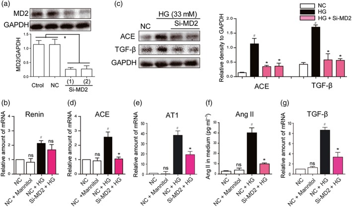
Figure 1.

MD2 silencing attenuated the activation of the RAS, induced by high glucose concentrations. (a) MD2 knockdown in NRK‐52E cells by siRNA approach. Two si‐RNA sequences for MD2 gene (Si‐MD2) were transfected in NRK‐52E cells and the MD2 protein levels were measured by western blot analysis (Ctrol: non‐transfected cells; NC: non‐MD2 scrambled transfection cells; two different siRNAs for MD2 were used). (b–g) Effects of MD2 knockdown in macrophages stimulated by high glucose concentrations (HG; 33 mM) (b, d, e, and g) Si‐RNA transfected or NC NRK‐52E cells were incubated with HG or 33‐mM mannitol for 12 hr. The mRNA levels of renin, ACE, AT1 receptors (AT1) and TGF‐β were detected by real‐time quantitative PCR assay with β‐actin used as housekeeping gene. (c and f) Si‐RNA transfected or NC NRK‐52E cells were incubated with HG or 33‐mM mannitol for 24 hr. Shown are representative western blot analysis and densitometric results for ACE and TGF‐β protein levels in whole cell lysate with GAPDH as a loading control. Ang II level in cultured medium was detected by ELISA kit. n = 5 independent experiments; bar graph shows mean values ± SEM; # P < .05, significantly different from NC group; *P < .05, significantly different from HG group; ns = not significantly different from NC + HG group
