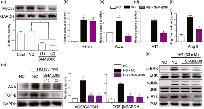
Figure 3.

MyD88 silencing and inhibition of MAPKs suppressed activation of RAS and expression of fibrotic factors, induced by high glucose concentrations. (a) MyD88 knockdown in NRK‐52E cells by siRNA approach. Two si‐RNA sequences for MyD88 gene (si‐MyD88) were transfected in NRK‐52E cells and the MyD88 protein levels were measured by western blot analysis (Ctrol: non‐transfected cells; NC: non‐MD2 scrambled transfection cells; two different siRNAs for MyD88 were used). (b–g) MyD88 silencing attenuated activation of RAS and MAPKs, induced by high glucose ((HG; 33 mM) concentrations. NRK‐52E cells were firstly transfected with si‐MyD88 and then stimulated by HG conditions for different times. (b–d) The mRNA levels of ACE, renin, and AT1 receptors (AT1) were detected by real‐time qPCR assay after 12‐hr incubation of HG. (e) ACE and TGF‐β were detected by western blot with GAPDH as a loading control after 24‐hr incubation of HG. (f) The Ang II levels in cultural medium were detected by ELISA after 24‐hr HG challenge. (g) The phosphorylation levels of MAPKs were detected by western blot analysis after 30‐min HG incubation. n = 5 independent experiments; bar graph shows mean values ± SEM; # P < .05 significantly different from NC group; *P < .05, significantly different; ns, not significantly different from HG group
