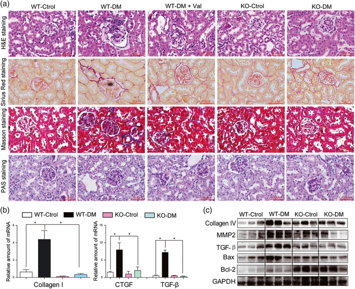
Figure 6.

MD2 knockout decreased diabetes‐induced fibrosis in mouse kidney. MD2 knockout mice (KO), valsartan (Val)‐treated C57BL/6 mice, and their wild‐type control (WT) developed diabetes (DM), 4 months after STZ injection. (a) Representative light micrograph of histochemical assessment of kidney tissues: haematoxylin and eosin (H&E) staining, PAS for glycogen (purple), Masson's trichrome stain (Blue) for detection of connective tissue, and Sirius red staining were used for the detection of fibrosis (Red); 400× magnification; scale bar: 50 μm. (b) Kidney tissue content of fibrotic genes was determined by real‐time quantitative‐PCR for collagen 1, CTGF, and TGF‐β. Bar graph shows mean values ± SEM; n = 8 mice per group, *P < .05, significantly different as indicated. (c) Representative western blot analysis of indicated proteins in kidney tissues
