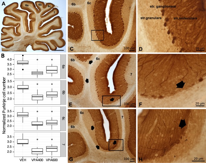
Figure 4.
Prenatal valproate exposure induced reductions in cerebellar Purkinje cell number. (A) Light micrograph of calbindin D28k (CB) immunostained sagittal section of rat cerebellar vermis at low magnification. (B) Boxplots representing normalized CB immunoreactive Purkinje cell numbers in cerebellar vermal lobules 6a, 6b, 6c and 7, for the VEH, VPA400 and VPA600 groups. Asterisks depict significant (p < 0.05) decreases compared to the VEH group. (C,D) Normal distribution of CB immunoreactive Purkinje cells in the ganglionic layer of cerebellar cortex on VEH sections. (E–H) Decreased CB-positive Purkinje cell number, found both in the VPA400 and VPA600 groups. The arrows point to unlikely large areas with lack of Purkinje cells. Scale bars: 1000 μm (A), 100 μm (C,E,G), 20 μm (D,F,H).

