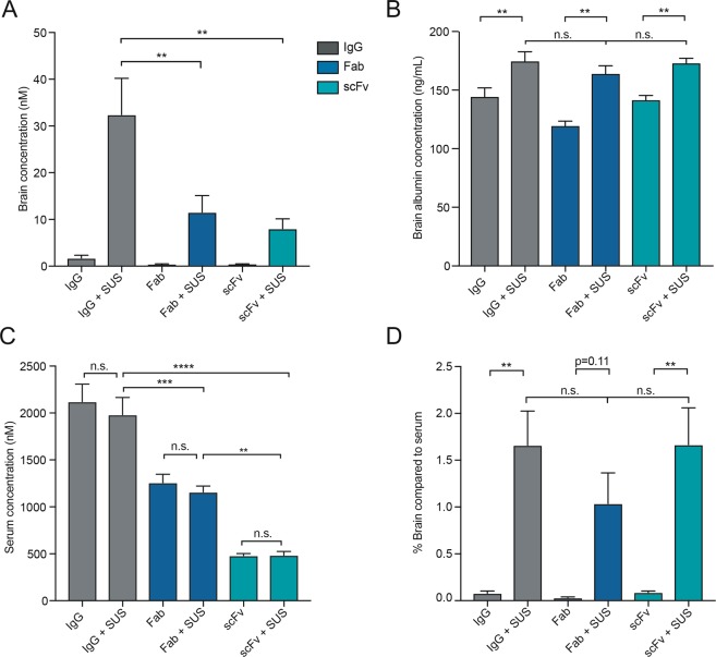
Figure 3.
Full-sized IgG antibodies reach a higher concentration in the brain following SUS treatment. (A) Determination of RN2N concentration in the brain post-delivery. SUS treatment increased the mean concentration of all formats in the brain (19-fold for IgG, 30-fold for Fab and 20-fold for scFv). Furthermore, following SUS, the concentration of the IgG was significantly increased compared to that of scFv and Fab. (B) SUS treatment increased the concentration of albumin in the brain, but there were no significant differences in albumin concentration in the brains treated with the different RN2N formats. (C) No significant difference in the serum concentration of RN2N was observed between mice treated with or without SUS. Serum concentrations of the IgGs and Fab formats were significantly higher than that of the scFv, in both the SUS- and sham-treated groups. (D) The percentage concentration of the antibody formats in the brain after SUS delivery compared to that in the serum was consistent between the different formats demonstrating that brain delivery following SUS was proportional to the corresponding serum levels. (n = 5, mean ± SEM; one-way ANOVA with Tukey’s multiple comparisons test; **P < 0.01, ***P < 0.001, ****P < 0.0001).

