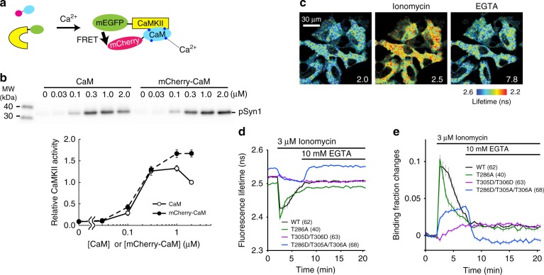
Fig. 1.
Design and characterization of CaMKIIα-CaM association sensor. a Design of a FRET sensor for CaMKIIα-CaM association. Monomeric EGFP (mEGFP) and monomeric Cherry (mCherry) fluorescent protein are fused to the N-terminus of CaMKIIα and the N-terminus of CaM, respectively. b mCherry-CaM activates CaMKIIα to the degree similar to non-labeled CaM at different concentrations of CaM in a cell-free system. Upper panel: western blot of phosphorylated Synapsin1 peptide (pSyn1) fused to mCherry. Lower panel: quantification of pSyn1 signal from 4 experiments, normalized with the pSyn1 signal at 2 µM non-labeled CaM. c Fluorescence lifetime images of CaMKIIα-CaM association sensor expressed in HeLa cells. d Time courses of fluorescence lifetime of CaMKIIα-CaM association sensor and its mutants (T286A, T305D/T306D and T286D/T305A/T306A) in response to bath application of ionomycin (3 µM) and EGTA (10 mM). e Time courses of changes in CaMKIIα-CaM association calculated from d. All data are shown in mean ± sem

