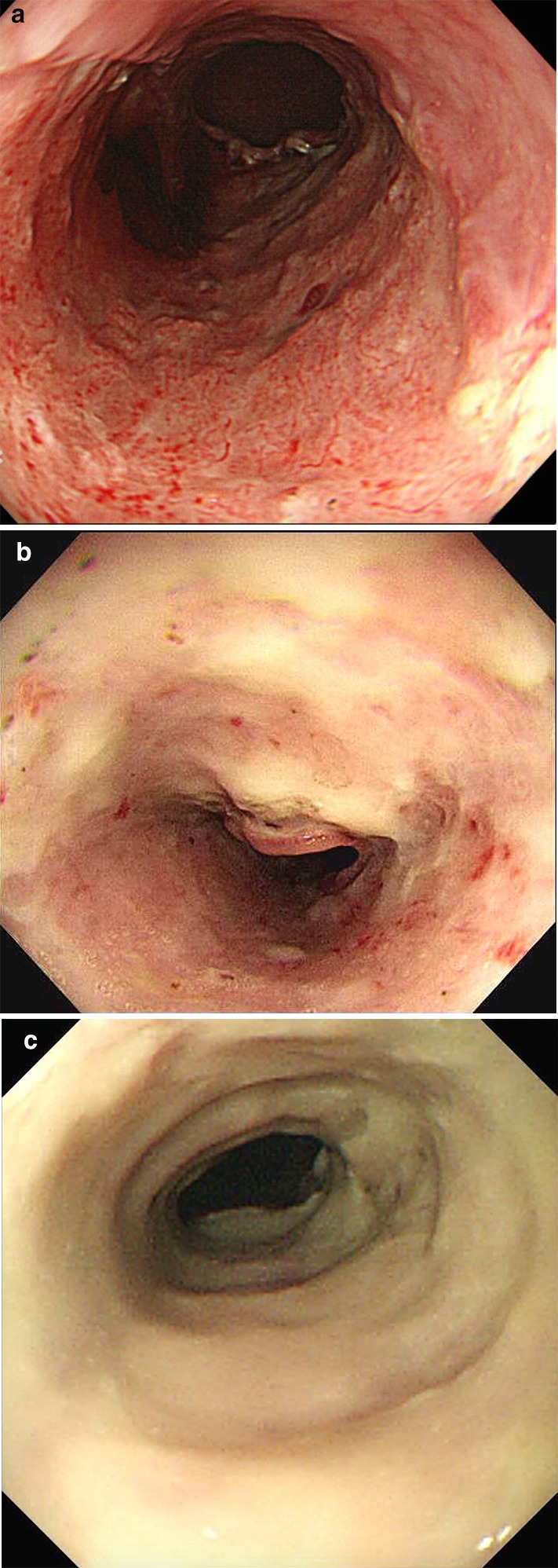
Fig. 1.

The white coats and blood vessels of the artificial ulcers after esophageal ESD. The groups were defined by visually according to the appearance of the artificial ulcer: in the thin white coat group, blood vessels were clearly visible (a); in the moderately thick white coat group, blood vessels that were partially visible (b); in the thick white coat group, no blood vessels were visible (c)
