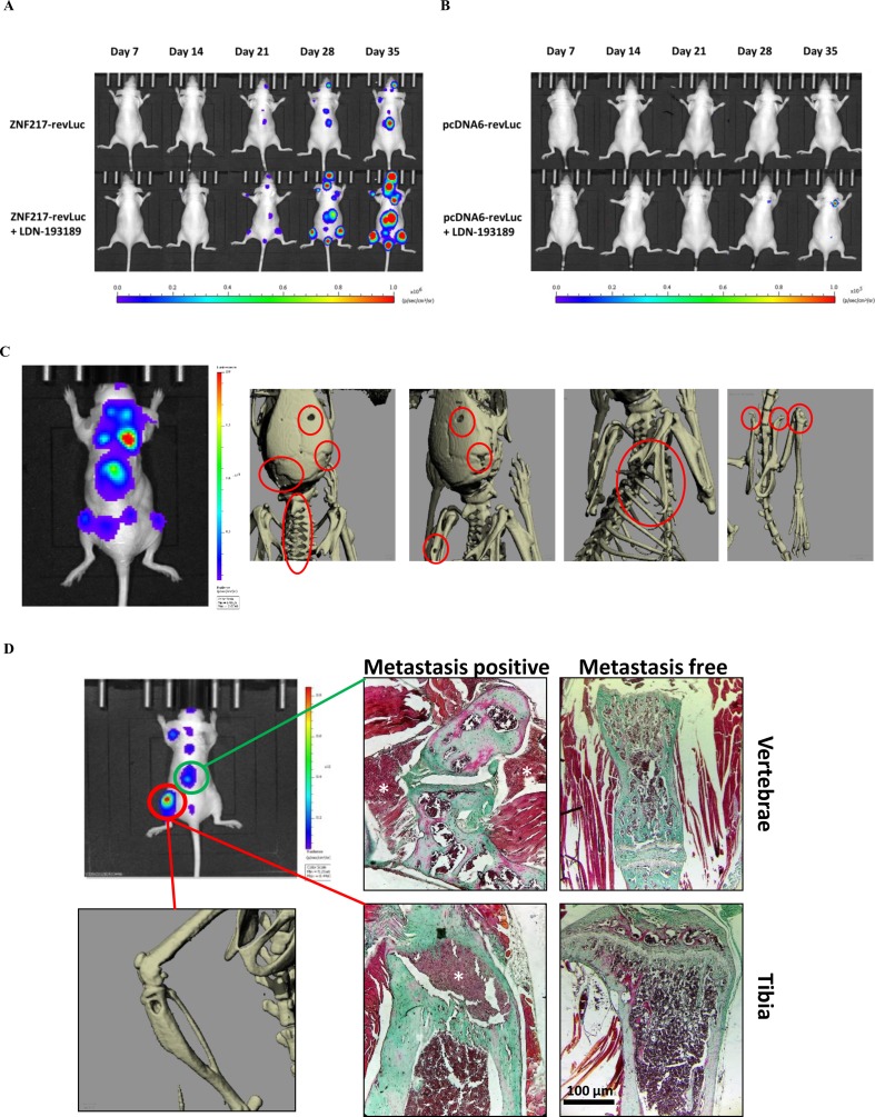
Figure 1.
Kinetics and development of metastases in vivo. The kinetics of metastases development were followed by bioluminescence in mice injected with (A) ZNF217-revLuc cells or (B) pcDNA6-revLuc cells, and treated or not with LDN-193189 (representation of one mouse per group representative of the entire group). (C) Whole-body bioluminescence and microCT imaging between 35 and 42 days after intracardiac injection of ZNF217-revLuc cells. (D) Representative bioluminescence image of an entire mouse exhibiting multiple metastasis foci including an osteolytic BM detected by microCT scan at the left tibia and by histological examination both at the vertebrae and the tibia from bone tissue sections stained with Goldner’s trichrome. Mineralized bone is stained in green and cells are stained in dark red. White stars identify metastasis foci. Histological examination of similar areas from a metastasis-free mouse is also shown in vertebrae and tibia. Scale bars: 100 µm.

