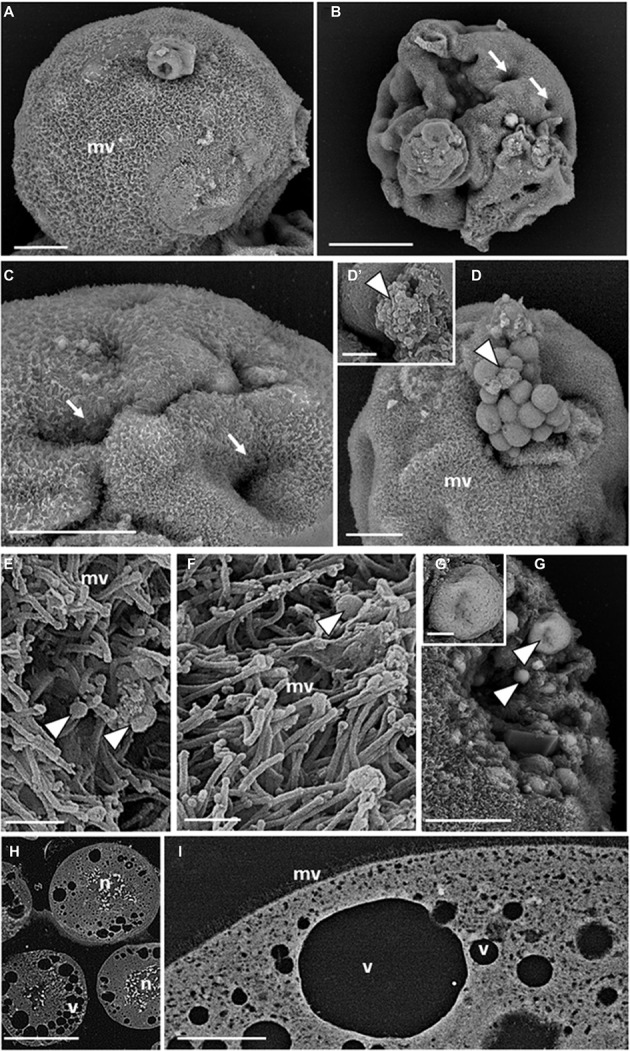
Figure 2.

SEM micrographs of teratocytes. Teratocytes isolated from pea aphids 5 days after parasitization by A. ervi have a surface covered with numerous microvilli (A) and are characterized by the presence of indentations or dimple-like structures (B,C). Spherical vesicles and structures similar to exosomes protrude from the plasma membrane [arrowheads in (D,D′)]. Numerous exosome-like structures [arrowheads in (E,F)] are interspersed among the microvilli. Spherical vesicles filled with smaller vesicles with circular profiles are visible in the cytoplasm [arrowheads in (G,G′)]. Large stellate nuclei (H) and vacuoles (H,I) are visible in the sectioned teratocytes. (mv) microvilli, (n) nucleus, (v) vacuoles. Bars in (A,C,D,G): 20 μm; bar in (B): 50 μm; bars in (D′,G′): 2.5 μm; bars in (E,F): 1 μm; bar in (H): 100 μm; bar in (I): 10 μm.
