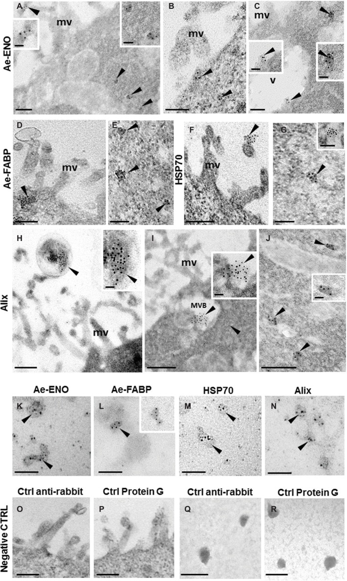
Figure 4.
Immunogold staining at TEM of Ae-ENO, Ae-FABP HSP70, and Alix. Gold nanoparticles (arrowheads) are visible in exosome-like vesicles released from teratocyte microvilli (A,F,H) underneath the plasma membrane (B–D), in the multivesicular body located next to the plasma membrane (I) and in those dispersed in the cytoplasm (A,C,E,G,J). Immunostaining assays performed on exosome-like structures released by teratocytes stimulated for 30′ with ATP 5 mM (K–N). No signal was detected in control experiments, where the primary antibodies were omitted, and samples were incubated only with the anti-rabbit secondary antibody (O,Q) or with the protein G gold-conjugated (P,R). (mv) microvilli; (MVB) multivesicular body; (v) vacuole. Bars in (A–G) and in (K–R): 100 nm; bars in (H–J): 200 nm; bars in inserts 40 nm.

