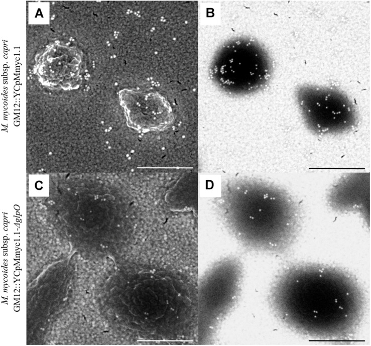
FIGURE 5.
Scanning electron micrographs showing immunogold labeling of the parental strain GM12::YCpMmyc1.1 (A,B) as well as the mutant strain GM12::YCpMmyc1.1-ΔglpO (C,D) incubated with anti-GlpO serum. Secondary electron micrographs show the cell surface of the mycoplasmas (A,C). Back-scattered electron micrographs reveal the presence of the gold-conjugated secondary antibody (B,D). Scale bar, 500 nm.

