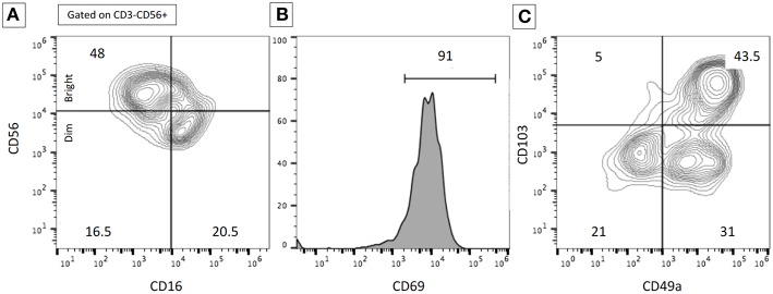
Figure 2.
Example of flow cytometry data illustrating the subset of resident lung NK cells. Flow cytometry analyses were performed on BALF in a patient with severe interstitial lung disease. The expression of the cell surface markers was performed after gating on CD3−CD56+ NK cells. (A) Proportions of CD56dim/bright and CD16+/− NK cells. (B) High expression of CD69+ on NK cells. (C) Proportions of resident NK cells according to CD103 and CD49a expression. The proportion of resident lung NK cells was higher than expected on normal lung samples. Numbers represent the % of the different populations.

