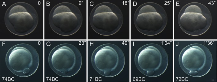
Figure 4.

Time‐lapse images of twin‐tail goldfish embryos. Individual time‐lapse images from a representative blastula‐period embryo (A–E) and gastrula‐period embryo (F–J). The embryos were incubated at 24°C. Lapsed times from the initiation of imaging are indicated at the upper right corner of each panel. Photos of embryos from fertilized eggs of Ryukin‐strain parents. Gastrula‐period embryos are indicated as blastopore closure (BC) in the lower left corner. Scale bar J = 0.1 mm. All embryos were photographed at the same magnification.
