
Figure 8.
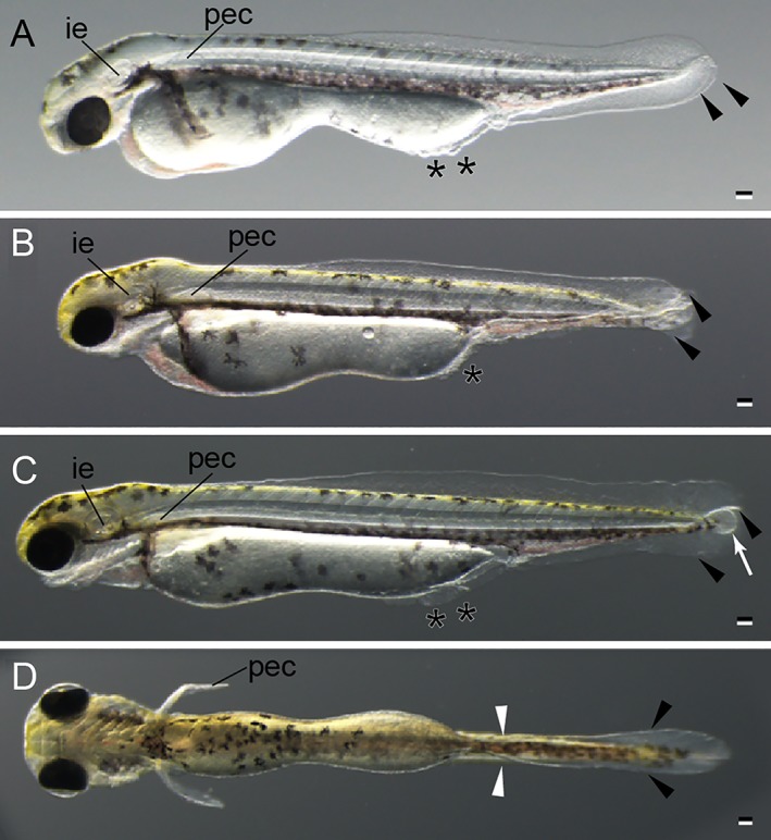
Hatching stage of twin‐tail goldfish larvae. A: Lateral view of long pec stage. B: Lateral view of pec fin stage. C: Lateral view of protruding‐mouth stage. D: Ventral view of protruding‐mouth stage. All larvae were derived from Ryukin‐strain parents. Black arrowheads and asterisks indicate bifurcated fin fold and malformed fin fold. White arrow indicates edema; white arrowheads indicate the edge of the bifurcated fin fold near the end of the yolk. ie, inner ear; pec, pectoral fin. Scale bars = 0.1 mm.