
Figure 9.
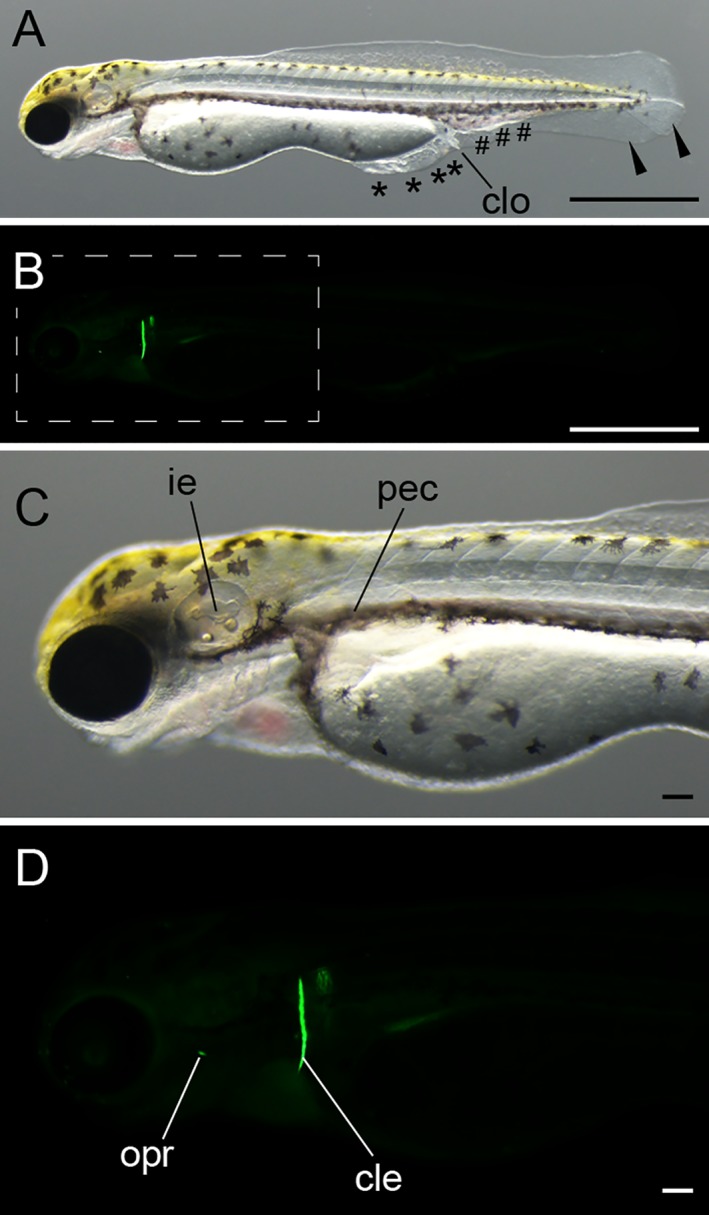
Protruding mouth‐stage larva. A: Lateral light microscopic view of the whole body. Black arrowheads indicate bifurcated fin fold; black asterisks indicate malformed fin fold; black pound signs (#) indicate enlarged blood island. B: Lateral view of calcein‐stained fluorescence. C: Magnified view of panel A. D: Magnified view of the boxed region in panel B. The pictured larva was derived from Ryukin parents. cle, cleithrum; clo, cloaca; ie, inner ear; opr, opercular pec, pectoral fin. Scale bars A,B = 1 mm. Scale bars C,D = 0.1 mm.