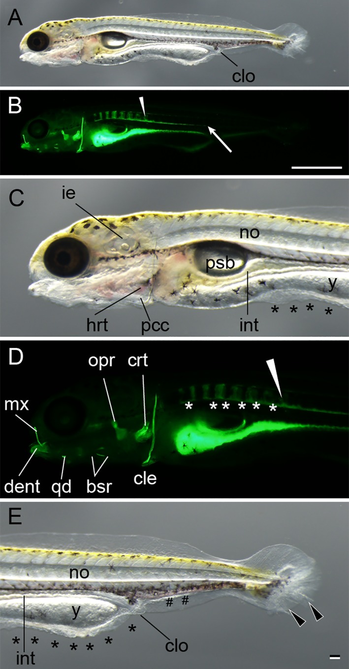
Figure 10.

Posterior swim bladder stage. A–E: Lateral views of a larva from Ryukin parents. Panels A–E are light microscopic views of the entire body, calcein‐stained fluorescence views of the entire body, magnified views of the anterior region of A, magnified views of the anterior region B, and magnified views of caudal region of A, respectively. Bifurcated caudal fins are indicated by the black arrowheads. Malformed fin folds are marked by black asterisks; enlarged blood vessels are marked by pound signs (#). White arrowheads mark the most posterior calcified vertebral body. White asterisks indicate calcein‐stained area between calcified vertebral elements. White arrow shows the posterior end of unsegmented calcein‐positive tissues on the ventral side of the notochord. bsr, branchiostegal rays; cle, cleithrum; clo, cloaca; crt, ceratobranchial; dent, dentary; hrt, heart; mx, maxilla; ie, inner ear; int, intestine; no, notochord; opr, opercular; pcc, pericardial cavity; psb, posterior swim bladder; qd, quadrate; y, yolk. Scale bar B = 1 mm. Scale bar E = 0.1 mm. Panels of the entire larva view (A,B), panels of the magnified view (C–E) were photographed at the same magnification.
