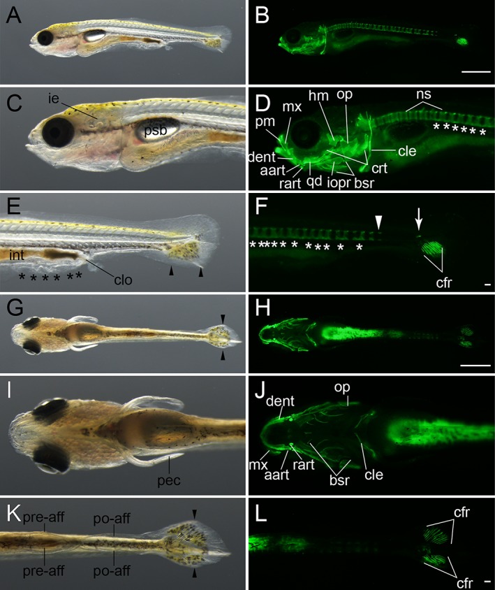
Figure 12.

Late caudal fin ray–stage larvae. A–F: Lateral views of an eight caudal fin ray–stage larva from Ryukin parents. G–L: Ventral view of an eleven caudal fin ray–stage Ryukin progeny. Left and right columns show light and calcein‐stained fluorescein microscopic images. Black asterisks and arrowheads indicate bifurcated caudal fin and malformed pre‐anal fin fold. White asterisks and arrowheads mark ectopically calcified notochordal region and the most posterior calcified centrum. White arrow shows calcified tissue at the level of the flexed notochord. aart, anguloarticular, bsr, branchiostegal rays; cfr, caudal fin rays; cle, cleithrum; clo, cloaca; crt, ceratobranchial; dent, dentary; hm, hyomandibular; ie, inner ear; int, intestine; iopr, interopercular; mx, maxilla; ns, neural spine; op, opercular; pec, pectoral fin; pm, premaxilla; po‐aff; post‐anal fin fold; pre‐aff, pre‐anal fin fold; psb, posterior swim bladder; qd, quadrate; rart, retroarticular. Scale bars B,H = 1 mm. Scale bars F,L = 0.1 mm. Panels of the first row (A,B), second and third rows (C–F), fourth row (G,H), and fifth and six rows (I–L) were photographed at the same magnifications.
