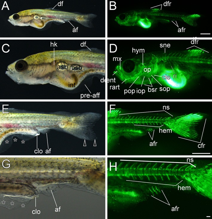
Figure 16.

Anal fin ray–stage larvae. A–H: Lateral views of three anal fin ray–stage larvae derived from Oranda strain. The left column (A,C,E,G) and right column (B,D,F,H) show light and calcein‐stained fluorescent microscopic images. Black arrowheads indicate bifurcated caudal fins. Black asterisks mark mutated area of pre‐anal fin fold. af, anal fin; afr, anal fin rays; asb, anterior swim bladder; bsr, branchiostegal rays; cfr, caudal fin rays; cle, cleithrum; clo, cloaca; dent, dentary; df, dorsal fin; dfr, dorsal fin rays; hem, hemal arch; hk, head kidney; hym, hyomandibular; int, intestine; iop, interopercular; mx, maxilla; ns, neural spine; op, opercular; pop, preopercular; pre‐aff, pre‐anal fin fold; psb, posterior swim bladder; rart, retroarticular; ri, rib; sne, supraneuralis; sop, subopercular. Scale bars B,F = 1 mm. Scale bar H = 0.1 mm. Panels at the first row (A,B), second and third rows (C–F), and forth row (G,H) are shown at the same magnification.
