
Figure 17.
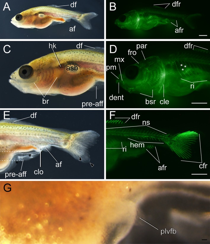
Lateral views of pelvic fin bud–stage larva. A–B: Whole‐body view of larva derived from Ryukin strain. C–F: Magnified views of anterior (C,D) and posterior (E,F) regions. G: Magnified view of the boxed area in E. Black arrowheads, black asterisks, and white asterisks indicate bifurcated caudal fin, mutated pre‐anal fin fold, and twisted part of ribs, respectively. af, anal fin; afr, anal fin ray; asb, anterior swim bladder; br, branchial; bsr, branchiostegal rays; cfr, caudal fin rays; cle, cleithrum; clo, cloaca; dent, dentary; df, dorsal; dfr, dorsal fin rays; fro, frontal; hem, hemal arch; hk, head kidney; mx, maxilla; ns, neural spine; par, parietal; plvfb, pelvic fin bud; pre‐aff, pre‐anal fin fold; pm, premaxilla; ri, rib. Scale bars B,D,F = 1 mm. Scale bar G = 0.1 mm. Panels at the first row (A,B), second row (C,D), and third row (E,F) are shown at the same magnification.