
Figure 7.
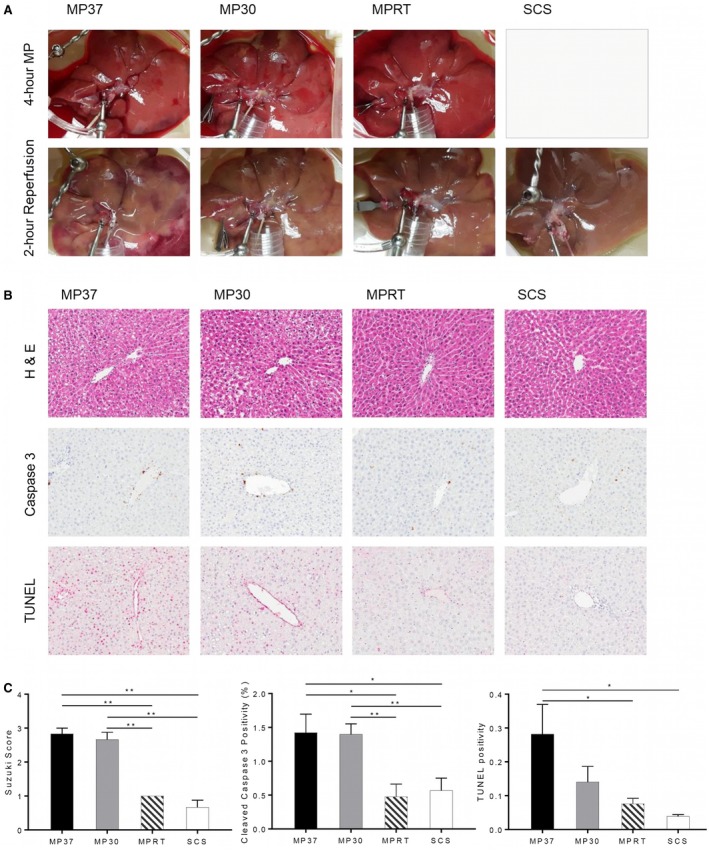
Histologic injury and apoptosis following reperfusion. Histology was performed at the end of graft reperfusion, including standard H & E and immunohistochemistry for cleaved caspase 3 and TUNEL. (A) Gross appearance of grafts during MP and reperfusion. (B) H & E and immunohistochemistry staining. (C) Quantitative analysis of graft injury and apoptosis by Suzuki score, cleaved caspase 3, and TUNEL staining. Data are shown as mean ± SEM, n = 6 per group for H & E, n = 5 per group for immunohistochemistry. *P < 0.05, **P < 0.01.