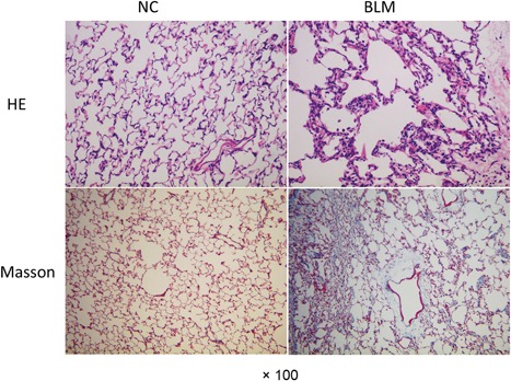
Figure 1.

Haemotoxylin and eosin (H&E) and Masson staining of lung tissues in rat models. Pulmonary fibrosis model rats were constructed and the fibrosis deposition was detected using H&E and Masson staining (×100). BLM, bleomycin group; NC, normal group
