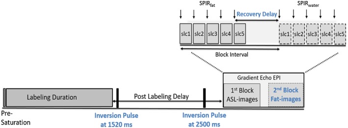
Figure 1.

Block diagram showing the multislice pCASL, including the fat‐navigator images and background suppression (setting: BGS2M), using a multiblock gradient echoplanar imaging readout. Detailed information about the two blocks of the readout is shown in the zoomed section (top right). In the first block ASL‐images (slc1–slc5) are acquired, each preceded by a SPIR pulse for fat suppression. After the SPIR pulse of the last slice (slc 5) fat recovery begins and the chosen recovery delay starts. pCASL, pseudo continuous arterial spin labeling; ASL, arterial spin labeling; EPI, echo planar imaging; SPIR, spectral presaturation with inversion recovery
