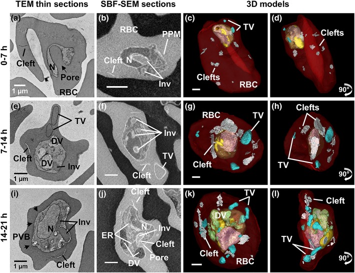
Figure 1.

Ultrastructure of P lasmodium knowlesi at ring and trophozoite stages. First column: transmission electron microscopy (TEM) of plastic‐embedded thin sections. Right hand columns: serial block‐face scanning electron microscopy (SBF‐SEM) images and rendered 3D models of the same cells. (a–d) 0 to 7‐hr postinvasion. (a) The arrow indicates the invagination pore. (b) The invagination (Inv) deforms the nucleus (N). (c–d) The early ring parasite occupies a small portion of the red blood cell (RBC). Small tubular vesicle (TV) and clefts are present in the RBC cytoplasm. (e–h) 7 to 14‐hr postinvasion. (e) A digestive vacuole (DV) is evident indicating initiation of haemoglobin digestion. (f) A branched invagination is observed. (g–h) Hemozoin crystals (green) are evident. Larger TV are observed. (i–l) 14 to 21‐hr postinvasion. Parasitophorous vacuole bulges (PVB) have formed (black arrows) and a pore in (j) indicates active haemoglobin uptake. (k–l) Larger DVs (green) accumulate next to the invaginations. Colours: PVM, pale yellow; nuclei, pink; invagination, yellow; DV, green; RBC, translucent red; clefts, white; TV, cyan. Panels on the far right represent 90o rotations of the preceding panels (see Videos S1–S6 for translations and rotations of these cells)
