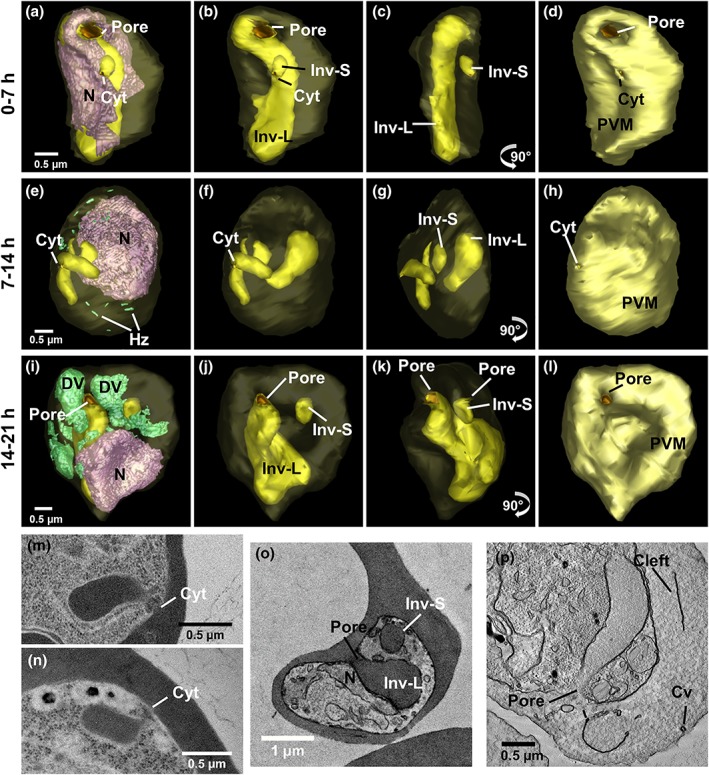
Figure 5.

Ultrastructural analysis of invagination features. Rendered serial block‐face scanning electron microscopy (SBF‐SEM) models of (a–d) early ring stage, (e–h) late ring stage, (i–l) and trophozoite stage. (m–n) Transmission electron microscopy (TEM) images of cytostomes in late ring/trophozoite stage. The cytostome is a collar‐like structure with a diameter of 80–100 nm (m,n). (o,p) Large pores (~400 nm) connect invaginations to the host red blood cell (RBC) cytoplasm. (p) Haemoglobin is depleted from the invagination upon equinatoxin II (EqtII) permeabilisation
