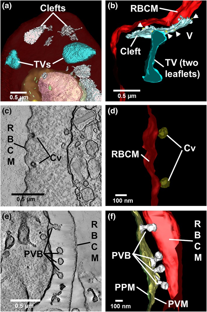
Figure 6.

Detailed views of rendered clefts and tubular vesicle (TV). (a,b) Rendered serial block‐face scanning electron microscopy (SBF‐SEM; a) and electron tomography (b) models showing Sinton Mulligan's clefts (white) and TV (cyan). Small vesicles (gold) appear to bud from the cleft. (c,d) Virtual section through (c) and rendered model of (d) caveolae (Cv, bronze) at the host RBC membrane. (e,f) Virtual section through (e) and model of (f) membrane blebs (rendered in white) forming at the PVM
