Figure 3.
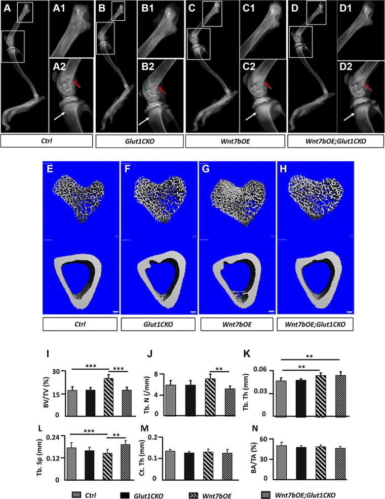
Inducible deletion of Glut1 diminishes Wnt7b-induced bone accrual. A–D2) Representative X-ray radiographs of the hind limbs. Boxed areas at a higher magnification (A1, A2, B1, B2, C1, C2, D1, D2). Red arrow, distal femur; white arrow, proximal tibia. E–H) μCT 3-dimensional reconstruction of trabecular bone (upper) from the proximal end or cortical bone (lower) from the midshaft of the tibia. Scale bars, 100 μm. I–N) Trabecular (I–L) and cortical (M, N) bone parameters from μCT analyses. BA, bone area; BV, bone volume; Ct. Th, cortical thickness; TA, total area; Tb. N, trabecular number; Tb. Sp, trabecular separation; Tb. Th, trabecular thickness; TV, tissue volume. **P < 0.01, ***P < 0.001, by 2-way ANOVA followed by Bonferroni’s post hoc test (n = 7).

