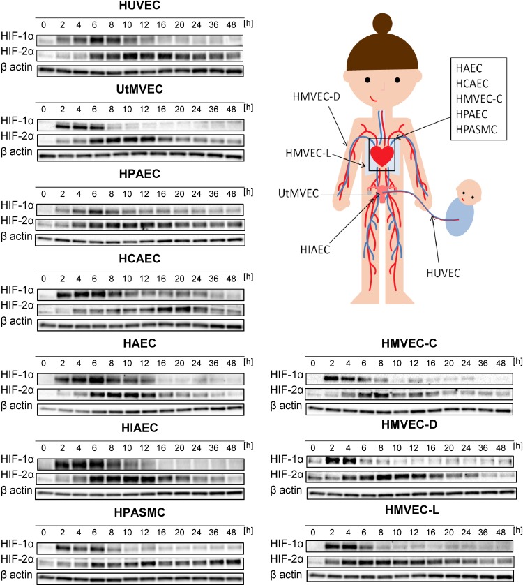
Figure 1.
Hypoxia results in accumulation of HIF-1α and HIF-2α in human ECs. Cells were exposed to hypoxia for the time periods specified, and total RNA and protein lysates were collected. The changes in HIF-1α and HIF-2α protein levels were evaluated by Western blot normalized to β-actin and total protein levels and related to the normoxic control. The densitometry analyses are provided in Supplemental Fig. S1. HIAEC, human iliac EC; HPAEC, human pulmonary artery EC.

