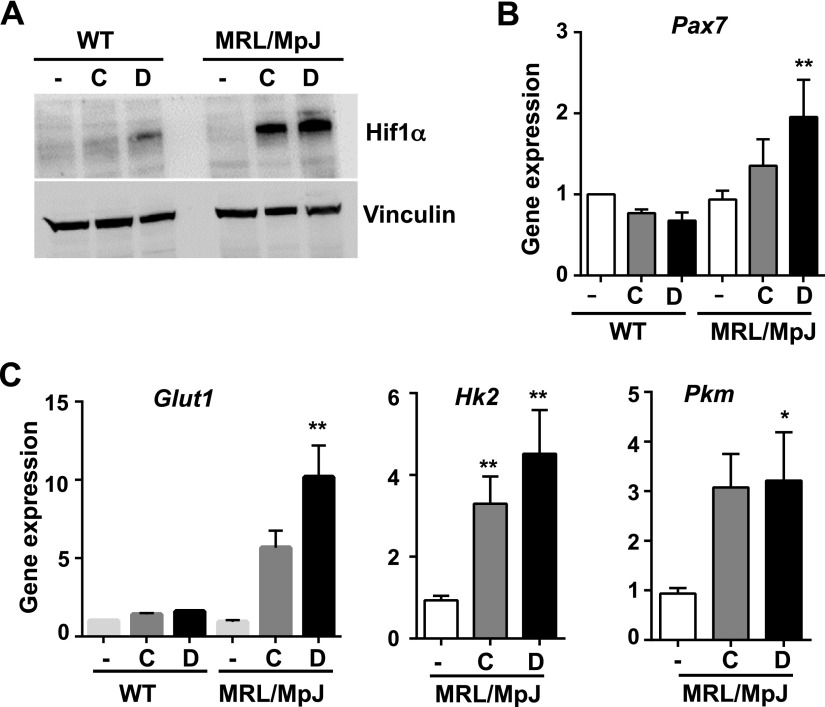
Figure 3.
Higher levels of HIF-1α and expression of glycolytic genes were found in MRL/MpJ MDSPCs than WT MDSPCs. A) Western blot analysis of the levels of HIF-1α in the lysates of WT MDSPCs and MRL/MpJ MDSPCs after treatment with DMOG (1 mM) and CoCl2 (200 μM) for 24 h. B, C) qRT-PCR for gene expression analysis in untreated and treated WT MDSPCs and MRL/MpJ MDSPCs for HIF-1α target genes, including Pax7, Glut1, Hk2, and Pkm. Error bars indicate means ± sem from triplicates (P < 0.05). *P < 0.01, **P < 0.001.

