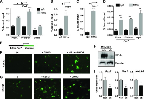
Figure 5.
ChIP assay for interactions of HIF-1α at its target genes in MRL/MpJ MDSPCs. Chromatin prepared from DMOG-treated MRL/MpJ MDSPCs was immunoprecipitated with control IgG and HIF-1α antibodies, followed by qPCR with gene-specific primers flanking HIF-1α responsive genomic sequence using ChIP DNA. A–C) Interactions of HIF-1α are indicated by % bound input: 3 regions in Pax7, the promoter region (Prom.), first intron, and 3′UTR as control region (A); the Vegfa promoter (B); and the Glut1 promoter that harbors HIF-1α –responsive genomic sequence and is indicated by a solid triangle (C). D) ChIP assay for HIF-1α occupancy at the Pax gene in CoCl2-treated MRL/MpJ MDPSCs. E) Schematic of a construct containing the 1.2-kb mouse Pax7 promoter driving the expression of Zsgreen (GFP). F, G) Fluorescent microscopic images taken after transient transfection with the Pax7-Zsgreen reporter gene in the myoblast cell line C2C12 (F) and HEK 293 cells (G), each in the presence of DMOG (1 mM) or CoCl2 (200 μM), and coexpression of mutant HIF-1α after transfection as indicated. Scale bars, 10 μm. H) Level of HIF-1α in MRL/MpJ MDSPCs after transducing with lenti-nshRNA and lenti-shRNA followed by precondition to hypoxic conditions for 24 h was analyzed with Western blot using the cell lysates. I) Quantitative gene expression showing mRNA levels of myogenic stem cell markers Pax7, Hes1, and Notch3 in nshRNA and HIF-1α shRNA MDSPCs from MRL/MpJ mice.

