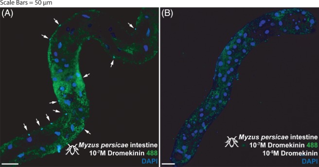
Figure 2.

Myzus persicae intestine (distal midgut and proximal hindgut) stained with 10−7 m kinin labelled with alexafluor488 (A) and then out‐competed with 10−5 m unlabeled kinin (B). (A) Staining apparent in a population of basal cells, characterized by overtly smaller nuclei (arrows). (B) Staining abrogated in basal (small nuclei) cells (realized by DAPI staining, arrows) during out‐competition with unlabeled 10−5 m kinin. Kinin‐F, green; DAPI, blue. Scale bars = 50 µm.
