Figure 4.

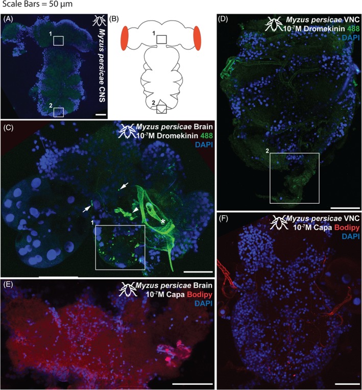
(A) Unstained Myzus persicae CNS, demonstrating baseline autofluorescent levels (488 nm excitation range; green). (B) cartoon schematic. (C) Myzus persicae brain and (D) VNC incubated with 10−7 m kinin‐F labelled with alexafluor488. (C) Staining apparent in a bilateral ‘ladder’ of neurons and a set of more baso‐lateral neurons in the suboesophageal ganglion (1white box). Position of 1white box indicated by 1boxes in (A) and (B). Staining also apparent in symmetrical pairs of neurons/neuronal clusters in the ventro‐ to dorso‐lateral protocerebrum (arrows). Some neurons obscured by cuticular material associated with the aphid feeding stylus (asterisk). (D) Little to no kinin‐F staining apparent in the VNC, although a faint set of cells in the most distal tip of the abdominal ganglion (2white box) are consistently observed. Position of 2white box indicated by 2boxes in (A) and (B). (E) Myzus persicae brain and (F) VNC incubated with 10−7 m CAPA‐1‐F labelled with TMR C5‐Maleimide. No apparent staining with CAPA‐1‐F in either brain or VNC. Kinin‐F, green; CAPA‐1‐F, red; DAPI, blue. Scale bars = 50 µm.
