Figure 4.

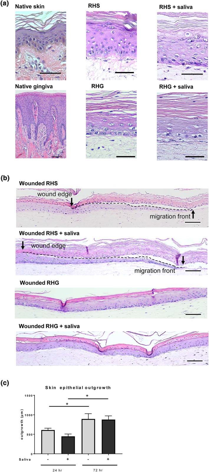
Blister wound healing model. (a) Representative haematoxylin and eosin staining is shown for native skin and native gingiva (left); reconstructed human skin (RHS; orthokeratinized epithelium) and reconstructed human gingiva (RHG; parakeratinized epitheliu; middle); RHS and RHG topically exposed to 100% saliva (right). (b) Freeze blisters were introduced into RHS and RHG. Re‐epithelialization is shown 72 hr after wounding with and without saliva treatment. RHS tissue sections showing original wound edge (black arrow), interface between dead epithelial layer and ingrowing epithelial layer (dotted line) and migrating front (arrow head). RHG tissue sections show that 72 hr post wounding, the fibroblasts have already migrated into the wound bed and the wound is completely closed, making it impossible to adequately define the exact wound margin. Scale bars represent 100 μm. (c) Quantification of re‐epithelialization after topical exposure to 0% (PBS) and 100% saliva for 72 hr, starting at time of introduction of freeze wound. * p < .05; ** p < .01; *** p < .001; two‐way ANOVA followed by Fisher's LSD test [Colour figure can be viewed at wileyonlinelibrary.com]
