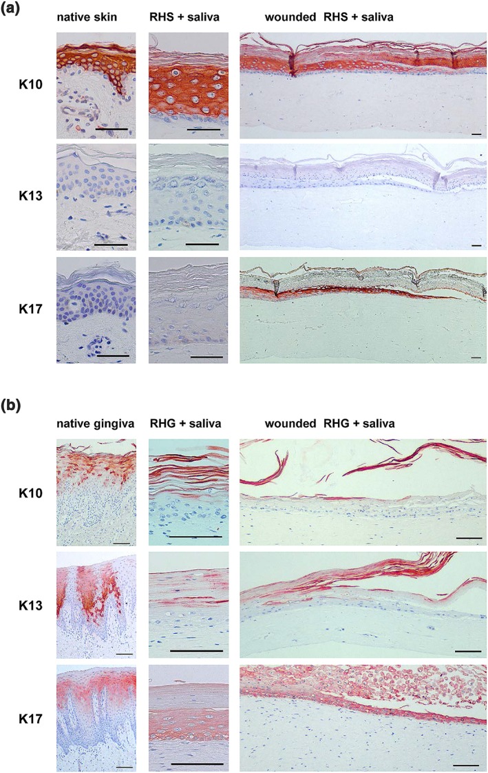
Figure 6.

Immunohistochemical staining with K10, K13, and K17 of (a) skin and (b) gingiva. Left: Healthy native skin or gingiva biopsy; middle: Unwounded area of RHS or RHG, which was exposed topically to 100% saliva for 72 hr; right: Epithelial migrating front of wounded RHS or RHG exposed topically to saliva for 72 hr. Scale bars represent 100 μm [Colour figure can be viewed at wileyonlinelibrary.com]
