Figure 1.

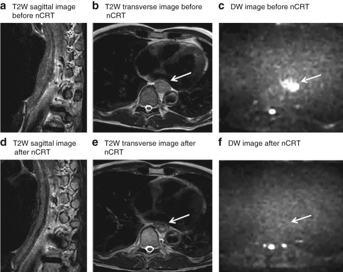
MRI of a patient with locally advanced oesophageal cancer that showed a pathological complete response to neoadjuvant chemoradiotherapy Images from a 55‐year‐old man with a cT3N0 lower oesophageal squamous cell carcinoma, and a complete pathological response after neoadjuvant chemoradiotherapy (nCRT) and oesophagectomy (tumour regression grade 1, ypT0 N0). a–c T2‐weighted (T2W) sagittal (a) and transverse (b) images before chemoradiotherapy show a hyperintense oesophageal wall, accompanied by a hyperintense signal on diffusion‐weighted (DW) imaging (c). d–f T2W sagittal (d) and transverse (e) images after nCRT show a hypointense oesophageal wall, indicating fibrosis; no high signal remained on the corresponding DW image (f). Arrows mark (initial) tumour location.
