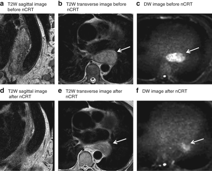
Figure 2.

MRI of a patient with locally advanced oesophageal cancer that showed pathological residual tumour after chemoradiotherapy and surgery Images from a 78‐year‐old man with a cT2 N0 lower oesophageal adenocarcinoma, who had residual tumour after neoadjuvant chemoradiotherapy (nCRT) and oesophagectomy (tumour regression grade 5, ypT2 N0). T2‐weighted (T2W) sagittal (a,d) and transverse (b,e) images before (a,b) and after (d,e) nCRT both show a hyperintense oesophageal wall. The corresponding b = 800 diffusion‐weighted (DW) images before (c) and after (f) nCRT demonstrate a clear hyperintense signal, highly suspicious for tumour. Arrows mark tumour location.
